Cell phone use has increased rapidly and public concern over the potential health effects of radiofrequency electromagnetic waves has increased. Therefore, the present study was conducted to evaluate the cytotoxicity in cell phone users by performing buccal cytome assay. Sixty male volunteers (20–25 years) using cell phones 3 to 9 hours per day were recruited with prior consent and performed buccal cytome assay in accordance with ethical guidelines. The individuals were divided into groups according to their addiction habits and call duration. The same number (n=60) of age-matched individuals who used cell phones for less than 1 hour and without any addiction were considered to be at lowest risk. The results of this study showed highly significant frequencies of various cell anomalies such as micronucleus, nuclear buds, pyknotic cells, karyotypic, condensed chromatin and karyolytic cells in at-risk individuals compared to low-risk individuals. Furthermore, the frequency of such cells was significantly higher in the group with maximum contact (7–9 hours) than in the groups with shorter call duration. Among exposed individuals, the addiction group showed a significant increase in these cell anomalies compared to the group without addiction. The results of our study indicate caution for cell phone users as they may be exposed to adverse long-term health effects, including cancer, from prolonged exposure to talking.
Introduction
Cell phones have become an indispensable device in our daily lives and emit radiofrequency electromagnetic waves (RF-EMW). These phones operate on frequencies depending on frequency usage in different countries, and are now used not only to talk, but also by growing children to access the Internet, data, pictures and videos. The increased use of cell phones over the past few years has exposed civilized individuals to RF-EMW, raising questions about health effects, particularly its long-term effects (WHO, 2006). Are. Some individuals are using cell phones to talk for long periods of time due to business need or long distance communication. Studies have previously suggested that chronic exposure to cell phone radiation leads to uncontrolled cell proliferation due to accumulated DNA damage and also that radio frequency electromagnetic waves (RF-EMW) exposure increases PKC (protein kinase C) activity. Reduces factors that may be associated with carcinogenesis (Desai et al., 2009). The use of cell phones, radar installations, and microwave ovens has increased around the world over the past several years, resulting in an alarmingly increased rate of humans being exposed to radio frequency waves.
Cell phones use microwaves as carrier waves in the frequency range between 300 MHz to 300 GHz. Aggarwal et al. (2008) have reported adverse effects of cell phones on semen including reduced sperm count, motility and morphology. Additionally, cell phone exposure has been linked to increased oxidative stress in semen which may impair male fertility (Agarwal et al., 2009). Researchers have attempted to link the much-discussed decline in human sperm quality over the past decade to increased exposure to RF-EMW, particularly through mobile phone use (Agarwal and Durairajanayagam, 2015). 140581038022208 The rate at which radiation is absorbed by the human body is called the specific absorption rate (SAR). The FCC (Federal Communications Commission) has set the maximum legal SAR of any handheld cell device at 1.6 watts/kg (Hamada et al., 2011 ) is limited to. The toxic effect of any chemical that has a harmful effect on the genetic material of living organisms is called genotoxicity.
Cell phone radiation is radiofrequency radiation that is classified under non-ionizing radiation, and therefore does not have the thermal effects responsible for the breaking of chemical bonds (Hamada et al., 2011). Several in vitro studies have reported evidence of genotoxic effects of RF-EMW, such as micronucleus assay (Koyama et al., 2003); Chromosomal aberrations (Garaj-Vrovac et al., 1991), DNA strand breaks (Diem et al., 2005). In contrast to the literature described above, some studies have reported negative results (Bisht et al., 2001; Speight et al., 2007). The WHO research agenda for radiofrequency fields has identified genotoxic endpoints as high priority research needs (WHO, 2006). In 2011 a group of international experts from IARC (International Agency for Research on Cancer) concluded that RF/MW (radiofrequency/magnetic waves) radiations should be listed as potentially carcinogenic to humans (Group 2B) ( Ban et al., 2011). This classification of RF as potentially carcinogenic to humans in Group 2B was not supported by the genotoxicity based mechanistic evidence given by Prihoda (2012). Although the data available so far is inconclusive, scientific evidence indicates some biological effects and potential adverse health effects that deserve further investigation.
A recent study observed DNA damage in oral cells after exposure to cell phone radiation, and concluded that mobile phone users may be prone to lethality and cytotoxicity (Gandhi et al., 2015). . Microwaves can have genotoxic effects on somatic cells of the human system and can also cause hereditary genotoxic effects in germ cells (Verschev, 2005). Another study on genetic polymorphisms of GSTM1 and GSTT1 in individuals exposed to radiation from mobile towers showed significant genetic damage (Gulati et al., 2015). The buccal micronucleus cytome (BMCyt) assay is a cost-effective, minimally invasive method to study DNA damage, chromosomal instability, cell death, and regenerative capacity of human buccal mucosa cells. It is now widely used in epidemiological studies to analyze the effects of nutrition, lifestyle factors, genotoxin exposure, DNA damage, chromosome segregation and cell death (Thomas et al., 2009). When using a cell phone for conversation, the placement pattern of the handset is by both ears and very close to the buccal cavity which maximizes exposure to RF-EMW radiations while talking. Therefore, this study was conducted to assess the cytotoxic effects in buccal cells of cell phone users and to correlate this effect on exposed individuals according to exposure time. Also, to find out the effects of various confounding factors like smoking, pan masala, chewing tobacco and alcohol consumption etc. on cell phone users.
Materials and Methods
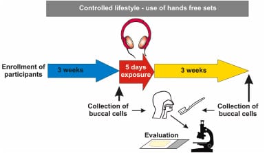
The study was approved by the institutional ethics committee and samples were collected in accordance with ethical guidelines and with the prior consent of cell phone users. A detailed questionnaire was filled out including the type of cell phone set, daily frequency of calls (incoming and outgoing), duration of use in 24 hours and years, specific absorption rate (SAR) of the model (obtained from the model’s website) and the brand in use. , Age, Occupation, Diet, Disease (if any), Addiction (if any), Allergy etc. The individuals at risk included 60 men who used cell phones 3-9 hours a day due to business needs or personal habits. The lowest risk group included an equal number of men (n=60). They were age-matched healthy individuals with low exposure to cell phone radiation (maximum up to 1 hour) and were completely non-smokers and had no other addictions.
Samples were collected from the inner walls of the cheeks using a sterile, small-headed plastic toothbrush, slides were prepared, and scoring was performed according to standard protocols (Thomas et al., 2009). The slides were stained with Giemsa, air dried and observed under a microscope. 1000 cells per subject were scored to determine the frequency of different cell types seen in the buccal cytome assay. Cells observed include normal cells, micronucleated cells (MN), binucleated cells (BN), nuclear buds (NB), pyknotic cells (PC), karyoblastic cells (KR), condensed chromatin cells (CC), and karyolytic cells (KL). Were. All data were expressed as mean ± standard error. Significance was considered when pp<o.o5. <mark id=”p_12″>Differences between the lowest and highest risk groups were analyzed using the Student test, while multiple comparisons between more than two groups (according to addiction habits and call duration) were performed by the Tukey multiple comparison test.
Results and discussion
Individuals at risk engaged in 20–100 calls per day due to job requirements, as many of them were in sales and marketing and had been using cell phones for 3 to 9 years and individuals at lowest risk engaged in 2–5 calls per day. Joined. SAR values of all models of handsets used by the study subjects were between 0.3-1.6 watts/kg body weight. In the present study 1000 normal buccal cells were scored and a variety of cell anomalies were observed. 140581038018112 The primary objective of this study was to evaluate the cytotoxic effects caused by cell phone radiation using the BMCyt assay because buccal cells are highly exposed to radiations while talking. Bisht et al. (2001) and Xellodar et al. Earlier studies on hematological parameters in serum samples have been reported by (2011). Aggarwal et al. (2009) showed harmful effects of cell phone radiation on sperm function and reactive oxygen species. Non-thermal DNA breakage in human fibroblasts as well as rat granulosa cells due to exposure to cell phone radiations is also reported by Diem et al. (2005).
Studies regarding the effects of cell phone radiation on oral cells have examined various parameters such as micronuclei (Hintsche and Storper, 2010; Yadav and Sharma, 2008); Have shown significant results with BN, MN, KR and KL (Rajkokila 2011). The present study is reporting for the first time the use of all cell types in the buccal cytome assay described by Thomas et al. (2009) as well as exposed individuals grouped according to exposure time and addiction. This is a direct in vivo study on the effect of cell phone radiation on oral cells. Our results showed a significant increase in Mn, Nb, PC, Kr, CC and Kl in exposed samples compared to minimally exposed individuals, and this increase was directly proportional to the duration of the call, as was observed in Group-4 (7–9 hours) compared with Group-2 (3-5 hours) and Group-3 (5-7 hours).
Among exposed individuals, group A (no addiction) and group B (addiction) showed highly significant increases in all cell anomalies (MN, BN, NB, PC, CC, KR and KL) compared to minimally exposed individuals . Furthermore, the addiction group showed a highly significant difference compared to the no-addiction group. In the exposed group, addictions to tobacco (26.6%), pan masala (16.6%), smoking (26.6%), alcohol (13.3%) were reported, while the remaining were non-addicted (16.9%). Celik et al. (2003) reported in their study of cytome assay on petrol pump attendants that cigarette smoking significantly increased the frequencies of micronuclei and other nuclear abnormalities in both control and exposed individuals.
The MN test in the BMCyt assay has been used to analyze genotoxic effects and monitor genetic damage in exposed individuals (Bonassi et al., 2011; Holland et al., 2008). Buccal cell MN has been identified as a useful biomarker related to oral cancer (Proya et al., 2006). Therefore, the frequency of MN in buccal mucosa cells can be used as a biomarker for genotoxic and carcinogenic agents, and the highly significant MN cells in our study may have serious consequences. Yadav and Sharma (2008) also reported similar results in buccal cells, while Hintzsche and Storer (2010) did not find significant results in their study of MN frequency in buccal mucosa cells of mobile phone users. In the present study, the frequencies of binucleated cells were found to be non-significant in exposed individuals compared to minimally exposed individuals, but were found to be significant when exposed individuals were further divided according to addiction habits and duration of calls.
The significance of the binucleated cells is unknown, but they probably indicate failed cytokinesis after the final nuclear division in the basal cell layer (Thomas et al., 2009). Nuclear bud (NBUD) is considered a biomarker of genotoxic events and chromosomal instability (Fenech et al., 2011). Cells with small shrunken nuclei and high density of nuclear material with intense uniform staining were identified as pyknotic cells (Tolbert et al., 1992). Karyoblastic cells were identified with a stronger presence of nuclear chromatin aggregation (compared to condensed chromatin cells). Cells with condensed chromatin, karyoblastic, pyknotic and karyolytic cells represent degenerated cells (apoptotic) (Holland et al., 2008). Since NBUD, pyknotic cells, condensed cells, and karyoblastic cells were significantly more observed in the exposed samples, it could be concluded that these cells were degenerating and were in early or late stages of apoptosis due to RF-EMW effects.
Cells appearing devoid of DNA and nuclei were identified as karyolytic cells, possibly indicating the final stages of cell death. In our study, karyolytic cells were found with significantly higher frequency in exposed individuals. Among exposed individuals, group-4 (7-9 hours) and adduct (group-B) showed maximum frequencies of karyolytic cells. Biomarkers found in the buccal micronucleus cytome assay such as pyknotic cells, nuclear buds, karyolytic cells, karyoblastic cells, etc. may be associated with several health risks and have proven to be a successful way to analyze cytotoxicity and genetic defects (Holland et al. ., 2008). Selappa et al. (2010) in individuals exposed to petrol observed increased MN frequencies in buccal mucosa cells that were associated with higher risk of cancer as a long-term effect and suggested careful monitoring. Some studies have shown that RF-EMW can induce oxidative stress in human saliva (Hamazani et al., 2013; Abu Khadra et al., 2015) while others reported no changes in oxidant/antioxidant profiles. (Khalil et al., 2014). Several reports showed no genotoxic effects due to RF exposure (Ros-Lore et al., 2012; Waldman et al., 2013). SA Derquist (2015) failed to show the significance of short-term exposure on biomarkers in volunteers exposed to cell phone radiations.
Some studies have shown that even at low levels RF-EMW can damage cell tissue and DNA, and have been linked to brain tumors (Hardell et al., 2007), cancer, impaired immune function, chronic allergic reactions, inflammatory reactions, (Jelloder et. A., 2011), has been linked to headaches, anxiety, stress, chronic fatigue syndrome and depression (Johansson, 2009). SA Derquist et al. (2012) No risk for parotid gland tumors due to short-term exposure was reported. Similarly, Daroit et al (2015) also concluded that, despite a significant increase in cell anomalies, the radiation emitted by cell phones among frequent users is within acceptable physiological limits. They also suggested further studies to investigate the harmful effects of cell phone radiation to draw a stronger conclusion. There is clearly ongoing debate about the potential harm to various organs of RF-EMW emitted by cell phones and growing public concern about its health risks.
In this study, among the exposed individuals, Group-B (addiction) and Group-4 (7-9 hours) had the least exposure followed by the least exposed individuals (Group-1), Group-A (non-addiction) and Group-2. (3-5 hours) and showed the highest level of DNA damage as compared to Group-3 (5-7 hours). Therefore, there is a strong correlation between cell anomalies and contact time, as maximum damage is observed in group-4 with the longest call duration (7-9 hours). Such increases may have long-term effects, including an increased risk of developing oral carcinoma in cell phone users after prolonged exposure (Fenech et al., 2011). Cell phone use has only increased in the last few years and all long-term effects may not yet be known.
Conclusion
The results of our study showed a highly significant increase in buccal cell anomalies including MN, BN, NB, PC, KR, CC and KL cells after exposure to cell phone radiations. Also, a strong correlation was observed between cell anomalies and cell phone exposure time. These cell discrepancies were further exacerbated by confounding factors seen in cell phone users with addiction (smoking, tobacco, and alcohol) when compared to a group without addiction. Abnormal cells seen in the BMCyt assay are used as an endpoint to detect cytotoxic damage in exposed individuals and this information may be helpful as an early warning of potential risk of genetic damage. Counseling and awareness of cell phone users becomes a necessity to protect them from the harmful effects of RF-EMW radiations.
Read Also:
- Effects of Mobile Radiations And Its Prevention
- Mobile Phone Radiations and Its Impact on Birds, Animals and Human Beings
- Development and Future Forecast of China’s Mobile Phone Industry
- Awareness Note On Mobile Tower Radiation & Its Impacts On Environment
- The Myth Of Cell Phone Radiation


Leave a Reply